These are a pair of glands located in the upper abdomen close to the spine and embedded in fat and loose connective tissue.
Each kidney is about 10 cm long, 6.5 cm wide, 5 cm thick, and weighs around 140 grams.

Vertical section through the kidney.
The outer margin of the kidney is convex; the inner is concave with a deep depression, known as the hilum, where the vessels enter and leave. The URETER, which conveys URINE to the URINARY BLADDER, also joins at this point. The ureter is spread out into an expanded, funnel-like end, known as the pelvis, which further divides up into little funnels known as the calyces. A vertical section through a kidney (see diagram) shows two distinct layers – an outer one, about 4 mm thick, known as the cortex; and an inner one, the medulla, lying closer to the hilum. The medulla consists of around a dozen pyramids arranged side by side, with their base on the cortex and their apex projecting into the calyces of the ureter. The apex of each pyramid is studded with tiny holes, which are the openings of the microscopic uriniferous tubes.
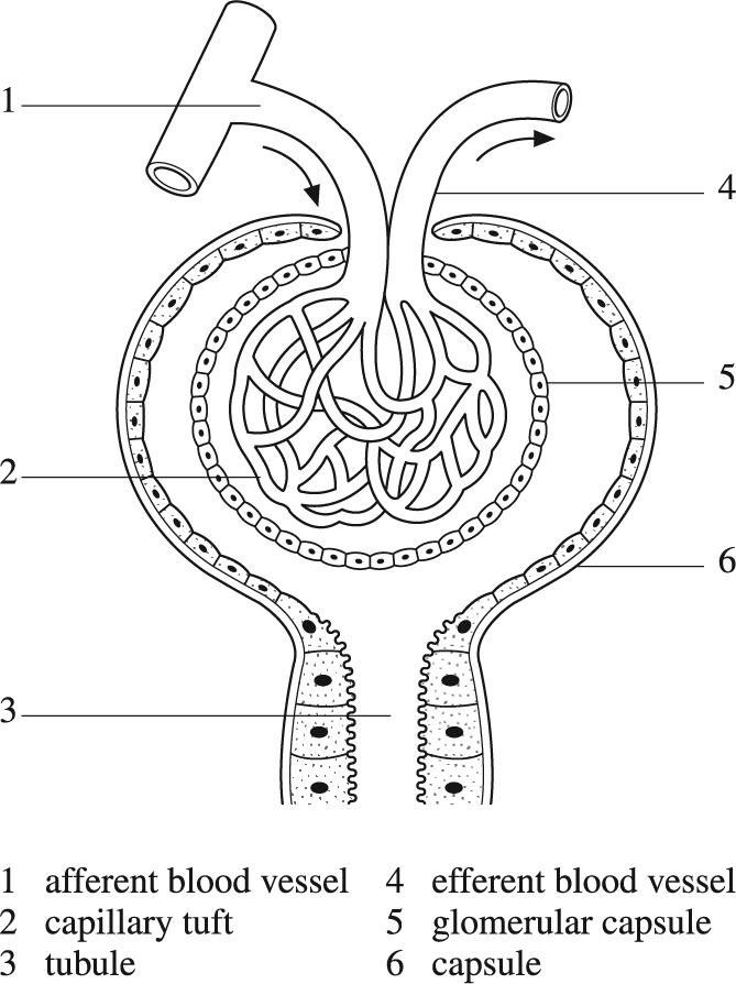
Diagram of glomerulus (Malpighian corpuscle).
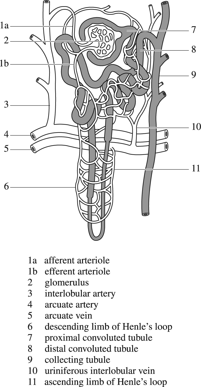
Diagram of renal tubules and blood supply of the kidney positioned in the cortex and medulla (see vertical-section diagram).
In effect, each pyramid, taken together with the portion of cortex lying along its base, is an independent mini-kidney. About 20 small tubes are on the surface of each pyramid; these, if traced up into its substance, repeatedly subdivide so as to form bundles of convoluted tubules, known as medullary rays, passing up towards the cortex. Each of these may be traced further back, and ends, after a tortuous course, in a small rounded body – the Malpighian corpuscle or glomerulus (see diagram). Each glomerulus and its convoluted tubule is known as a nephron and constitutes the functional unit of the kidney. Each kidney contains around a million nephrons.
After entering the kidney, the renal artery divides into branches, forming arches where the cortex and medulla join. Small vessels come off these arches and run up through the cortex, giving off small branches in each direction. These end in a tuft of capillaries, enclosed in Bowman's capsule, which forms the end of the uriniferous tubules just described; capillaries with capsule constitute a glomerulus.
After circulating in the glomerulus, the blood leaves by a small vein, which again divides into capillaries on the walls of the uriniferous tubules. From these it is finally collected into the renal veins and leaves the kidney. This double circulation (first through the glomerulus and then around the tubule) allows a large volume of fluid to be removed from the blood in the glomerulus, the concentrated blood passing on to the uriniferous tubule for removal of certain salts, etc. Other arteries come straight from the arches and supply the medulla direct; the blood from these passes through another set of capillaries and finally into the renal veins. This circulation is confined purely to the kidney, although small connections by both arteries and veins exist which pass through the capsule and, joining the lumbar vessels, communicate directly with the aorta.
The function of the kidneys is to separate fluid and certain solids from the blood. The glomeruli filter from the blood the non-protein portion of the plasma – around 150–200 litres in 24 hours – 99 per cent of which is reabsorbed on passing through the convoluted tubules.
Three main groups of substances are classified according to their extent of uptake by the tubules:
These include amino acids, glucose, sodium, potassium, calcium, magnesium and chlorine (for more information, see under separate entries).
only when their concentration in the filtrate exceeds that in the PLASMA, such as UREA, URIC ACID and phosphates.
from the tubular fluid, such as CREATINE; these accumulate in kidney failure, resulting in general ‘poisoning’ known as URAEMIA.